Blood analysis of unvaxed blood samples
Is there nanotechnology in people's blood after exposure to vaccinated?
I have the wonderful pleasure being able to us an Echo Revolve microscope, thank-you very much Dr. Henry Ealy and the AGES confernce group! I will periodically post some images and videos of live blood analysis. I am still not entirely sure what I am seeing in some of these samples, feel free to comment or share ideas. I have been following others that are also looking at blood samples. I hope that my work can add to the research being done.
This first video clip shows blood from an unvaxed person that has not had any recent exposure to vaccinated people. You can see healthy, round, red blood cells, a couple white blood cells, no fibers forming between the red blood cells, no rouleaux formation, and no moving dots or other unusual things in this sample. The white blood cells look a little strange but this person had recently had a cold and seems to have more white blood cells than should normally be present.
This video shows an unvaxed blood sample after this person had exposure to vaccinated people for approximately 4 hours. You can see that the red blood cells are stacked in the rouleaux formation and there are dots moving around in a weird random pattern. The dots are approximately 1-4 microns in size, compared to the red blood cells that are approximately 8 microns in diameter. (I had to video the ipad screen with my phone so I appologize for the light relfections).
This is the same sample a few minutes later and at higher magnification. You can now see that fibers are forming between the red blood cells and you can more clearly see the moving dots and fibers between the red blood cells. I do not see the randomly moving dots in healthy blood samples.
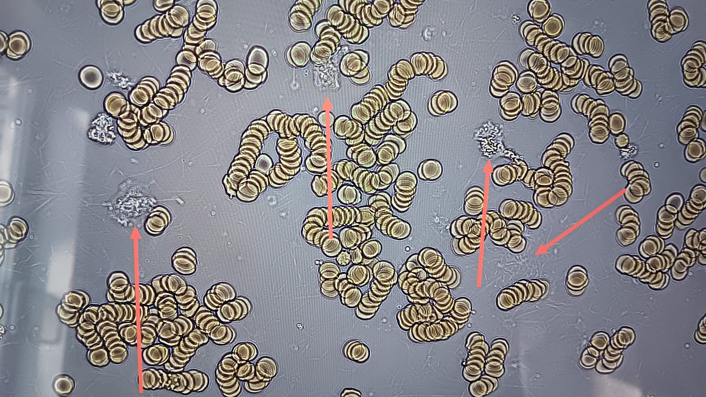
I noticed that in the unvaxed with exposure, the blood looks almost normal immediately after the blood is taken (by finger prick and then drop of blood dropped onto the slide and coverslip on top, takes approx 30 seconds to drip the blood and then visualize under the microscope), the blood then starts to form more rouleaux and then about 3 minutes later fibers start appearing between the red blood cells. It appears that the dots are moving around these fibers. The image below is taken within 1 minute of the blood sample taken. There are some white blood cells with the red arrows that look normal and there are no fibers yet.
The image below is the same place on the slide, red blood cells clumping together, fibers forming, and white blood cells look unhealthy. My guess is that the fibers are fibrin, only because I looked at a blood sample of a person taking aspirin and they had rouleaux and moving dots but no fibers. I will get a video of that soon.
Here is the same image but zoomed in a bit more.
I plan to continue with blood analysis and I will look at all varieties of blood samples with and without exposure, will include vaccinated blood, look at blood samples after people have tried various supplements, and I will figure out how to get images from the microscope so I don’t have to take pictures of the screen with my phone :)
Dr. Wendi


